


The ankle joint is often injured because it bears heavy loads. A doctor can diagnose ankle arthrosis based on symptoms and prescribe treatment. The disease does not depend on age and sex, tissues become thin and destroyed, which can lead to disability.
12% of the population suffers from arthrosis, and it is more common among women of retirement age.
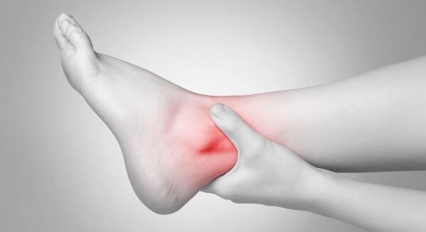
As mentioned, the ankle joint can withstand a lot of stress. It keeps the body upright and allows the person to move. Its violation changes the usual way of life.
Osteoarthritis of the ankle joint (symptoms and treatment may vary) is a chronic disease in which irreversible processes occur in the cartilage.
The disease occurs gradually. A healthy person has a smooth joint surface. When overloaded, it promotes easy sliding during physical activity.
In arthrosis, tissue nutrition and metabolism deteriorate. The outer surface of the joint changes, becomes rough, the cartilage touches and inflammation appears. When a person lifts heavy objects, the load falls on the bone, which leads to degenerative disorders.
If treatment is not started, severe pathologies develop. In later stages, cartilage and tissue are affected, the synovial membrane is irritated, and the joint loses stability. In this case, the supporting function suffers, movements become difficult.
Species
There are several types of arthrosis depending on different criteria:
- causes of occurrence (primary, secondary);
- stages of arthrosis;
- localization of pathology;
- forms of localization (generalized and local);
- disease course (acute and chronic).
| Classification criteria | Types of arthrosis |
|---|---|
| place of manifestation | arthrosis of the knee, wrist, ankle, elbow, shoulder and cervical arthrosis. |
| cause of origin |
|
| localization |
|
| the course of the disease |
|
Ankle arthrosis is divided into primary (degenerative processes begin in healthy cartilage due to excessive physical activity) and secondary (destructions are diagnosed, dystrophic changes appear in the cartilage tissue).
Phases and degrees
Arthrosis of the ankle joint (symptoms and treatment directly depend on the age of the patient) can appear in different ways. In some, many years pass from the appearance of the first symptoms to the critical stage, while in others the disease develops rapidly.
It depends on the age and accompanying diseases when the therapy started. As ankle arthrosis progresses, the symptoms become more pronounced.
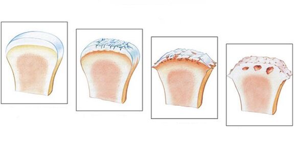
There are 4 stages of the disease.
- The first stage is often overlooked. Main symptoms: stiffness that occurs in the morning, characteristic creaking when walking. Pathogenic changes are not detected in the image, the destructive process has already begun.
- Stiffness lasts longer in the morning. It will take 20-30 minutes for the leg to develop. Some patients have lameness. On the x-ray, you can see stage 2 pathology by bony growths and bone displacement.
- In stage 3, symptoms become more pronounced. Painful sensations appear in a calm state, the patient cannot do without painkillers. Lameness becomes noticeable and sometimes crutches are needed. The joint swells, changes, muscles become thinner and decrease in volume. The joint space narrows, which can be seen on an X-ray, and osteophytes form.
- The last stage develops in the absence of treatment. Cartilage is destroyed, joint surfaces grow together. The patient cannot walk.
There are several degrees of arthrosis:
- First degree– X-ray shows no changes or articulations. There is a slight morning stiffness. At this stage, it is necessary to start treatment.
- In the second degreeactivity becomes difficult, you hear creaking when walking, swelling is observed. X-ray shows a reduction of the interarticular space. The person limps and the morning stiffness lasts longer.
- In the third degreeclearly expressed cruciarthrosis, joint deformation. Muscles atrophy even more, movements become limited. Constant rest is required. The pain does not go away even in this state.
- On the last stepThere is practically no common space, activity is almost impossible. X-ray allows you to diagnose a large number of osteophytes. Only surgical intervention is prescribed.
Osteoarthritis of the ankle joint occurs gradually, so treatment should be started when the first symptoms appear in order to prevent worsening of the condition and emergence of complications.
Symptoms
Osteoarthritis of the ankle joint is characterized by several symptoms (influence on the method of treatment):
- The pain is initially moderate and occurs only during physical activity. Over time, the pain becomes stronger and bothers you at rest;
- in the case of injuries and dislocations, swelling and inflammatory manifestations appear, and the temperature increases in the area of the injury;
- "dry" click accompanied by pain;
- dislocation, as the cartilage becomes thinner and degrades, the joint loses stability. Bones move and fall out of the joint capsule;
- stiffness of the joints;
- when walking, the person gets tired quickly;
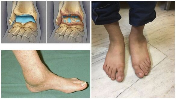
- in the last stages, the joint is deformed.
If at least one symptom appears, consult a doctor immediately.
Reasons for occurrence
Osteoarthritis of the ankle joint (symptoms and treatment are often caused by age changes) affects the older generation. Recently, pathology has been observed among young people.
The provoking factors are:
- injuries, dislocations and bruises;
- age-related disorders in joints and ligaments;
- inflammatory processes;
- overweight;
- violation of metabolic processes;
- congenital foot deformity and flat feet that occurred during life;
- hereditary predisposition;
- excessive physical activity;
- using uncomfortable shoes;
- diseases of the endocrine system;
- osteochondrosis.

Synovial fluid is produced less, which is why the cartilage is less nourished. The joint space narrows, which can lead to bone fusion. Crusarthrosis appears, which cannot be reversed. Nevertheless, treatment should be prescribed immediately to prevent the progression of the disease.
Diagnostics
The diagnosis of arthrosis consists of the study of existing symptoms and data obtained from research. Since there are no tests that can clearly determine the pathology, doctors recognize laboratory methods as insufficiently effective.
During remission, the indicators are normal, and during relapse, the blood test shows an increased level of ESR and c-reactive protein. This means that the pathology has already begun.
Instrumental methods are used to confirm the diagnosis:
- Simpleradiographyis the most reliable method. Muscles do not perceive X-rays equally: soft ones transmit them, and hard ones absorb them. The study reveals the disease itself and its consequences.
The image allows you to analyze the condition of the bone surfaces in the joint, the shape, size and position of the structures in relation to each other, the condition of the tissues and the size of the joint space. Thanks to this data, the degree of pathology can be determined.
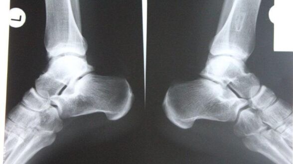
If the ankle joint is involved, the diagnosis is made in lateral, posterior, and posterior projections with the foot moved inwards. If there are appropriate symptoms (narrowing of the joint space, osteophytes and other signs), arthrosis is diagnosed.
- Nuclear magnetic resonancedetermines the disruption of the functioning of hydrogen molecules under the influence of a strong magnetic field. It allows you to explore parts of the body that contain water.
The dark shade in the image represents the bones, as their water content is much lower, while the muscles, nerves and discs appear lighter. Diagnostics also reveals minor disorders in bone tissue and joints. The procedure is indicated before joint replacement. The only drawback is the high price of diagnostics.
- M. R. Ivery precisely examines the ligamentous structure of the joint, muscle tissue and cartilage. Thanks to the study, the expert can assess the condition of the lower leg joints, which enables identification of the pathology at the very beginning of its development. The procedure is painless and takes about 30 minutes.
During the procedure, the person is affected by radio waves and strong magnetic radiation. It must be remembered that the magnetic field is dangerous for the physiological state. MRI is prohibited in case of neuropsychiatric disorders, pregnancy and the presence of metal objects in the body.
- Ultrasoundenables accurate diagnosis. The device produces waves that reflect off the tissue and are recorded on a screen. The doctor examines the picture and makes a diagnosis. For the clarity of the image, a gel is used that removes air and ensures easy movement on the surface.
The advantages of this procedure are health safety, affordable price and high precision.
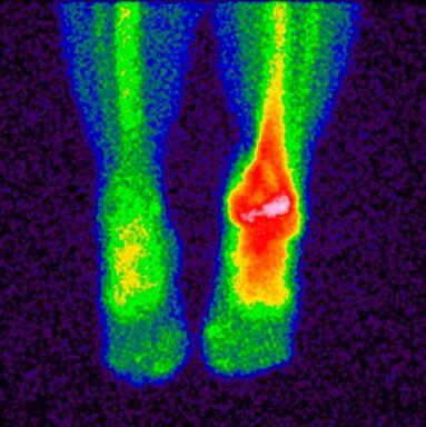
- Bone scintigraphy- a study that enables the determination of pathological disorders in the bones using isotopes. A special substance containing labeled atoms is injected into the patient's body. Pathological areas are divided into cold and hot.
In the first, there are no isotopes, the blood flow to them is weaker, and they are not detected during the scan. This includes places where malignant tumors have appeared. In hot areas, isotopes collect more actively and are clearly detected during scanning. These areas indicate the appearance of inflammatory processes.
This study makes it possible to separate arthrosis from similar diseases with similar clinical signs, and based on the results, the doctor gives a prognosis and prescribes treatment.
The main contraindications for the study are carrying a child, breastfeeding and taking drugs containing barium.
- Wrist punctureis a procedure in which the doctor inserts a needle into the joint cavity to collect synovial fluid for analysis.
This biomaterial continues to be studied in the future, and based on the results, the specialist determines the characteristic features of the disease and at what stage of development it is. In case of arthrosis of the ankle joint, a puncture is performed in the front part between the outer ankle joint and the tendon of the long extensor of the fingers.
When to see a doctor
If the treatment of arthrosis is not started on time, there is an inability to work, and sometimes even disability. Some patients are in no hurry to seek help because they do not know which doctor to make an appointment with. At the first symptoms, it is necessary to visit a rheumatologist who diagnoses dystrophic and inflammatory changes in the joint.
You should contact him if:
- there is discomfort and pain in the joints after excessive load, at the end of the working day;
- it is difficult to find a comfortable position for the legs at night;
- joints swell, skin turns red;
- there is a sharp pain, it is difficult to move;
- squeaking and clicking sounds appear;
- the joints are deformed.
With the help of modern diagnostic and therapeutic techniques, it is possible to avoid surgical intervention and preserve the functioning of the joint.
Prevention
Osteoarthritis of the ankle joint (symptoms and treatment can be checked with a doctor) can be prevented.
In order to prevent arthrosis, experts recommend following certain rules:
- wear comfortable, well-fitting shoes without heels;
- maintain a proper diet, drink enough clean water;
- choose a suitable vitamin-mineral complex;
- exercise;
- walk more often in the fresh air;
- avoid excessive load on the legs;
- avoid hypothermia;
- be regularly under the supervision of a doctor;
- give up bad habits;
- do a set of ankle warm-up exercises.

It is especially important to adjust your diet. Nutritionists have agreed on a menu that will prevent the disease from worsening and saturate the body with the necessary substances.
- You must eat often and in small portions.
- Drink at least 2 liters of clean water.
- Avoid sweet and salty foods.
- Do not eat 4 hours before going to bed.
- Steam, bake, cook food.
Fasting and a strict diet in arthrosis are strictly prohibited in order to prevent the leaching of calcium necessary for the restoration of bones and cartilage.
Treatment methods
Once the diagnosis is confirmed, treatment must be started immediately. It is impossible to completely get rid of arthrosis, the main thing is to slow down the destructive processes and increase the period of remission. Various techniques are used for this purpose.
Medicines
Different drugs are used to treat arthrosis:
- Anti-inflammatoryand painkillers eliminate the source of inflammation and relieve pain. Tablets and ointments are used. The sooner anti-inflammatory drugs are taken, the greater the chance of saving the joint.
- Glucocorticoidsthey are used if the above drugs do not give the desired result. They are produced in the form of a solution for injections and injected into the joint.
- Chondroprotectorsnecessary to slow down the process of cartilage destruction.
The treatment regimen and dosage of drugs is compiled by the doctor based on the severity of the symptoms, accompanying diseases and other factors. It is strictly forbidden to self-medicate in order not to worsen the situation.
Traditional methods
As for traditional methods of treating arthrosis, doctors recognize their beneficial properties and positive effects. Traditional medicine is also used as disease prevention.
The main prescriptions for the treatment of arthrosis of the ankle joint are as follows:
- Burdock leaves are washed well and applied with the soft side to the skin. The plant is fixed with a bandage or transparent film and left overnight.
- Heat sea salt (buckwheat, sand) in a pan, pour it into a linen cloth and apply it to the painful area. Keep until the salt cools down. This is an effective way to relieve pain.
- Cover the lilac with triple cologne, leave in a dark place for 2 weeks, rub the sore spot twice a day.
- Grind eggshells into powder, take 0. 5 tsp. before eating.

The use of traditional treatment methods must be agreed with the doctor. This is not the only measure, but an addition to the main therapy.
Other methods
When conservative therapy does not bring positive effects, they resort to radical measures - surgery.
As a rule, indications for surgery are:
- repeated and primary arthrosis grade 3-4;
- complications;
- strong and long-lasting pain radiating to the knee;
- apparent lameness;
- paralysis of leg muscles;
- deterioration of joint flexion-extension properties and the ability to support the foot.
The following surgical interventions are used for foot arthrosis:
- Arthrodesis– joint immobilization surgery. Its task is to restore the lost ability to support the limb. The main disadvantage is the probability of bone fusion, which leads to immobility, so it is used very rarely.
- Arthroscopyis a minimally invasive procedure in which the doctor cuts the joint and inserts an arthroscope. The surgeon performs a visual examination and assesses the state of the intra-articular structures, and if necessary, removes parts of the damaged joint or blood clots from the synovial fluid. With this operation, the risk of recurrence is too high.
- Endoprostheticsit is carried out in particularly difficult cases. It allows replacement of a damaged joint in a certain part or completely. Prostheses with modernized mechanics are used and last up to 20 years.
The main contraindications for surgery are age up to 12 years, fistulas in the joint, diabetes mellitus, cardiac dysfunction and infectious diseases.
Possible complications
If treatment is delayed or missed, the following complications may occur:
- disability;
- irreversible deformation;
- joint inactivity and immobility;
- decline in quality and standard of living.
In addition to these complications, the chronic course of the disease is accompanied by pain, discomfort and the inability to lead an active lifestyle.
In order for gymnastics, medicines and folk treatment to be more effective, it is recommended to use special orthopedic devices that reduce the load on the joint. This includes an orthosis and a fixation bandage.
The orthosis fully follows the contours of the ankle joint, increases the range of motion, relieves swelling and pain. A fixation bandage has the same effect as an orthosis. It is made of soft elastic fabric that allows you to secure the joint well. The bandage is used only in periods of remission, when the exacerbation has passed.
Osteoarthritis of the ankle joint is a serious disease that, if not completely treated, leads to severe consequences and complete immobility of the joint. Diagnosis in the initial phase, careful attention to symptoms and competent therapy make it possible to avoid surgical intervention.